
PhD thesis defense to be held on March 29, 2024, at 11:00 (virtually)
Picture Credit: Kostakis Gkiatis
Thesis title: Presurgical human brain mapping with magnetic resonance imaging
Abstract: The critical requirement to preserve basic human functions after brain surgery has given rise to the need for functional imaging of these processes pre-operatively to map them in the brain and avoid their extraction. Motor, speech, memory and vision are just some of the functions that can be mapped preoperatively. In the early stages of functional imaging, electroencephalography flourished. However, it quickly became inadequate methodology for imaging more complex functions such as speech. The invasive intracarotid amygdala technique was extensively used and evaluated to find the hemisphere of speech lateralization and is now considered the gold standard for evaluating other techniques. Its invasive nature, however, has made neurophysiologists look for alternative techniques. In the modern era, functional magnetic resonance imaging has gained ground in preoperative evaluation, with most centers worldwide (96%) having the ability to perform the technique, while in contrast, the intracarotid amygdala technique has declined with only 76% of centers having the ability to perform it. In Greece, the centers that have functional magnetic resonance imaging are few in number. In addition to this, there are no language mapping protocols for functional magnetic resonance imaging that have been validated for their validity and their ability to successfully map the language network reliably in all the subjects.
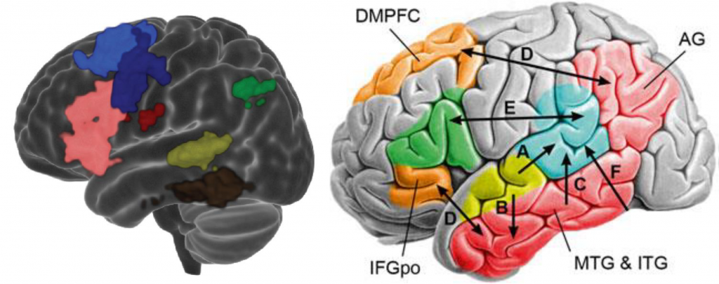
In this PhD thesis, we seek to confirm the reliability and validity even in the subject-level analysis of a protocol for language mapping in Greek. For this objective, a protocol that has already been confirmed in the literature in English was translated and transferred into Greek language and culture. Twenty healthy Greek native speakers were volunteered to perform these tasks to identify the regions expected to be activated in the healthy population. Data acquisition was performed at " St. Luke" hospital, Thessaloniki, Greece. All six critical language areas were found activated, Broca's area, Wernicke's area, Exner's area, angular gyrus, supplementary motor area and basal temporal language area. Additional activations were found in the hemisphere of the cerebellum opposite to the hemisphere of language lateralization, as well as some clusters in the thalamus, the caudate nucleus and the hippocampus.
Following that, in this PhD thesis, we seek an alternative technique to the statistical analysis technique of the general linear model. The trigger in this direction is the inability of the general linear model to satisfactorily map the activated areas in subjects who are either unable to cooperate during the tasks or unable to remain still, causing noise to interfere with brain activations making the results unreliable. The alternative technique studied in this thesis is the independent component analysis which can extract the sources that compose the original signal of functional magnetic resonance imaging. We show that the independent component analysis is able to produce reliable results in 89.59% of the scans while the general linear model was able to produce reliable results in only 88.03% of the scans. More importantly, however, is that the combination of both techniques is able to produce reliable results in 92.66% of scans improving the sensitivity of functional magnetic resonance imaging for language mapping.
Supervisor: Professor George Matsopoulos
PhD Student: Kostakis Gkiatis